Fetal cells retrieved from fluid in the uterus can now be turned into balls of cells called organoids, which could help diagnose and treat fetuses with a serious lung condition
By Clare Wilson
4 March 2024
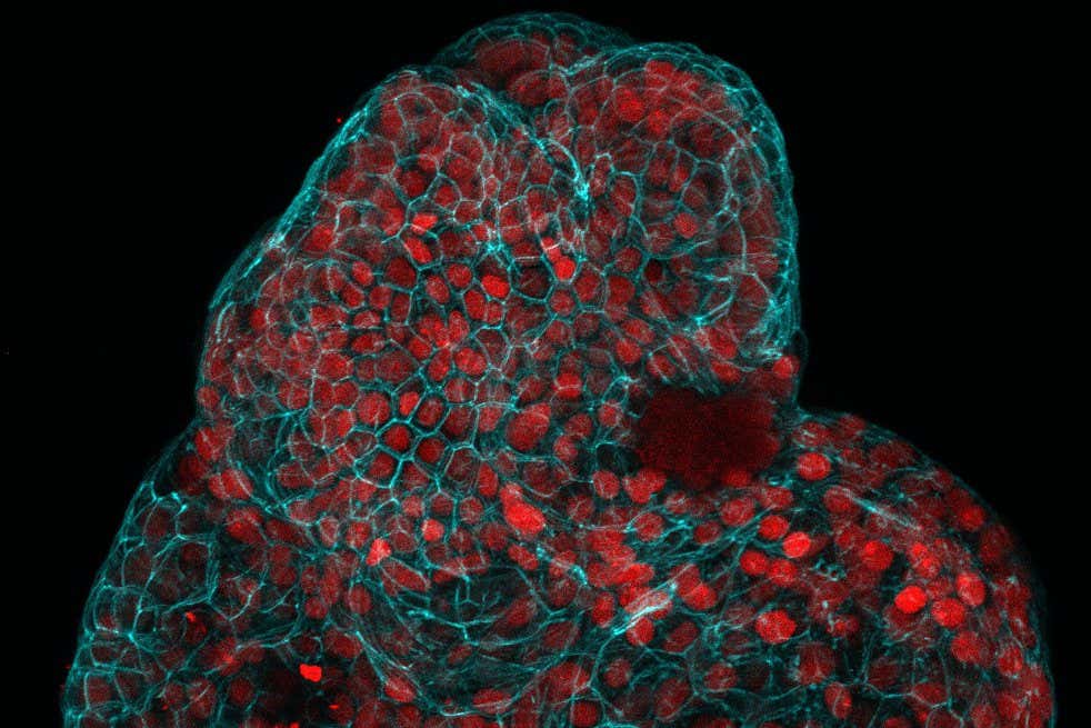
Balls of cells grown from amniotic fluid. The red indicates lung stem cells
Giuseppe Cala, Paolo di Coppi, Mattia Gerli
Babies born with serious medical conditions could one day get better diagnoses and treatments while in the uterus, thanks to a new technique that involves taking samples of cells from fluid in the uterus and growing them in a dish.
In a world first, Paolo De Coppi at Great Ormond Street Hospital in London and his colleagues have shown that fetal cells from amniotic fluid can be coaxed into forming miniature balls of lung, kidney or small intestinal tissue. They also showed these lung organoids could potentially help guide the treatment of babies born with a sometimes-fatal lung condition called congenital diaphragmatic hernia (CDH).
The technique hasn’t yet been used to treat any children, but the results show that is possible in principle, says De Coppi. It could also be modified to help in various other congenital conditions in a strategy the researchers call “personalised prenatal medicine”.
Advertisement
Read more
Unravelling the secrets of the vagus nerve will revolutionise medicine
The idea exploits a recent approach in which cells in a dish are coaxed to grow into tissue organoids, about the size of a lentil, which take up a three-dimensional structure. These then capture certain aspects of the tissue in question, including whether it is healthy or growing abnormally, better than the standard technique of growing cells in a two-dimensional layer.
The team has now shown that samples of amniotic fluid taken during pregnancy contain fetal cells capable of forming organoids of tissue from the lungs, kidneys and small intestine.